This is an assortment of cases done right in our office! Our patients have all consented to have their photos published here for the benefit of readers. To satisfy the very curious and dentally knowledgable, some more detail about each case has been included.
Minor Concerns
Restorations
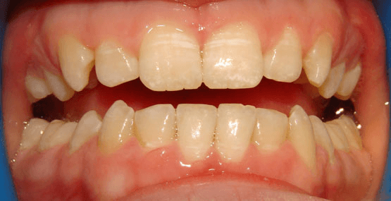
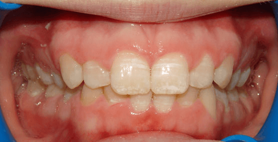
The main issue here was not decay or fracture, it was the appearance of the upper canine teeth (upper eye teeth – third ones from the middle). In the first photo, their horn-like appearance is quite obvious. Instead of shaving away any tooth material, conservatively bonding resin material to the teeth eliminated the undesirable appearance. What’s nice about newer white filling materials is their chameleon-like abilities to pick up colour from the teeth. (H3PO4 (aq) etch, Kerr Optibond FL adhesive only, Dentsply Esthet-X A2 resin. Microabrasion was done later to address the upper incisor dysmineralization)
Microabrasion
Microabrasion is a great, non-invasive way to remove congenital superficial brown and white spots on teeth. This underutilized technique employs a gently abrasive slurry that removes discolourations without compromising tooth strength or decay resistance. When cases are selected properly, they can often produce results better than doing fillings or even porcelain veneers. The dark brown stains often end up with the best results, which are visible immediately! No local anesthetic is required for this service.
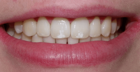
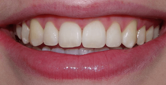
This patient disliked the white patches visible on front teeth. A single session of Microabrasion resulted in immediate improvement, with anticipated further improvement as time progresses. (Ultradent Opalustre 6.6% HCl Microabrasion slurry)
Whitening
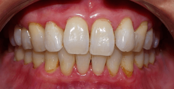
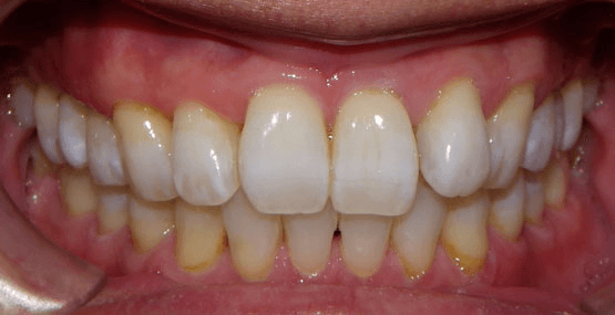
This is Dr. Wong’s own mouth! Pre and post-whitening photos using KoR-Night Whitening gel in custom trays for two weeks. (KoR-Night 16% carbamide peroxide gel from Evolve Dental Technologies)
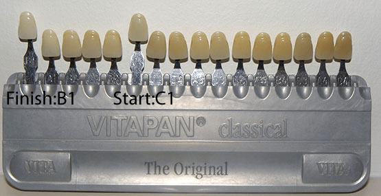
Photography often doesn’t accurately show changes in whitening, so we’ve included the shade guide that gives you a better idea:
Dr. Wong’s teeth went from a C1 to a B1 – a change of 5 shades brighter. (Vitapan Classical shade guide, arranged in order of value, or brightness).
Moderate Concerns
Veneers and Crowns
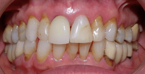
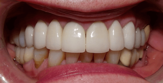
This is a longstanding patient who had not done anything about her smile for a while. She had come faithfully for her hygiene appointments and was cavity-free, but finally admitted that she wanted to do something about her teeth. She disliked the mismatched colours of her teeth, as well as the upper tooth crookedness and unevenness. If you look closely, one of her upper front teeth was already a crown, but in order to redo her smile it was necessary to replace it with one that would match the other teeth that would be done.
Seven crowns (on teeth 14,12,11,21,22,24,25) and two veneers (on teeth 13,23) , all e.max lithium disilicate from Ivoclar Vivadent. Teeth were air abraded and acid etched using Bisco’s Uni-etch 32% H3PO4 (aq) with BAC. Crowns were cemented with Unicem2 (3M) and veneers were bonded in with Calibra Adhesive Resin cement as per recommended protocol. Lab work courtesy of Image Dental Laboratory in Barrie.
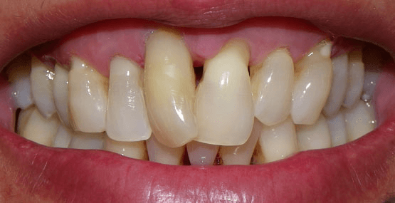
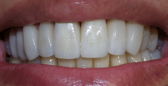
This patient was also a long-time patient of our office, coming regularly for years but always hating the very rotated upper right front tooth, and general unevenness of upper and lower front teeth. We decided to place two crowns and six veneers on the upper front teeth, as well as one crown and two veneers on the lower front teeth. Given the extreme rotation of the upper front tooth as well as the patient’s refusal of any orthodontic straightening, we first had to complete a root canal on that tooth. All restorations e.max lithium disilicate from Ivoclar Vivadent. Crowns on teeth 11,21,32 and veneers on teeth 15,14,13,12, 22, 23, 31, 41.
Teeth were air abraded, acid etched using Bisco’s Uni-etch 32% H3PO4 (aq) with BAC, and finally bonded in with Calibra Adhesive Resin cement as per recommended protocol. Lab work courtesy of Image Dental Laboratory in Barrie.
Smile Reconstruction
Case 1
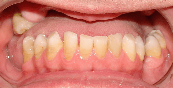
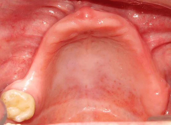
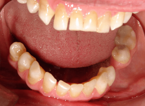
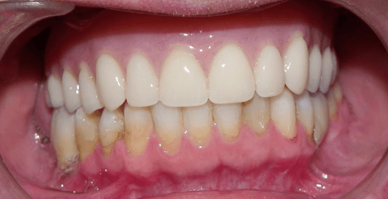
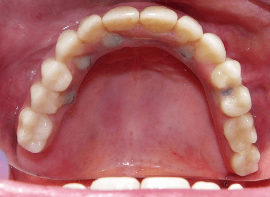
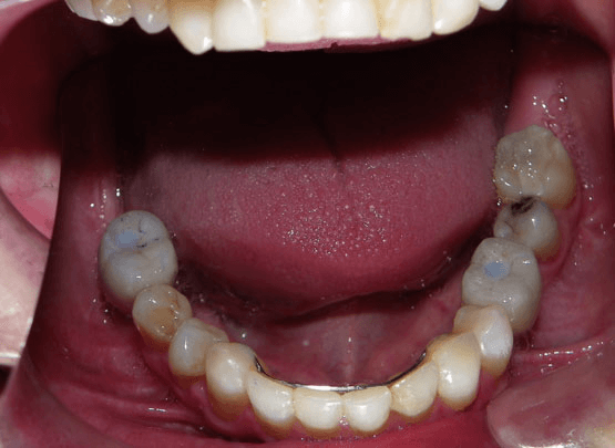
This patient had been wearing a complete upper denture from a young age, and had also lost many lower teeth. He was having difficulty chewing due to a combination of denture loosening as well as missing teeth, and decided that he would like a complete set of teeth that did not need to be removed from the mouth for cleaning. On the upper jaw, bone atrophy over the years required significant bone grafting.
Ultimately, a complete arch of teeth was placed, anchored in the mouth by six implants. On the lower jaw, drifting of teeth required preliminary orthodontic treatment (fixed braces) to optimize tooth positions and close spaces. Following that, implants were placed on both sides to replace the missing teeth. The collapse of his bite has been corrected and the patient can now eat and smile without fear of a denture coming out.
(Maxilla: bone grafting was done bilaterally from bone harvested from the patient’s hip. Six Nobel Biocare Replace implants were placed (four 4.3mm, two 5.0mm) and a fixed upper denture supported by a NobelBiocare Titanium CAD/CAM-milled framework was used as the final prosthesis. Mandible: Fixed orthodontic treatment to close anterior spacing as well as upright premolars. Tooth 45 was left rotated due to significant preexisting rotation. Large natural space between 45 and 44. Bilateral nerve lateralization surgery in the premolar areas followed by placement of one 4.3mm NobelReplace implant per side. Oral surgery courtesy of Dr. Cameron Clokie, Profiles Oral Surgery, Toronto. Orthodontics and prosthetics by Dr. Elston Wong.)
Case 2
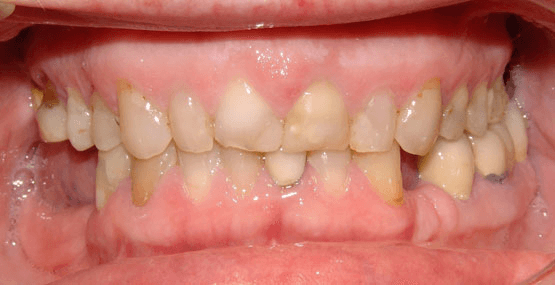
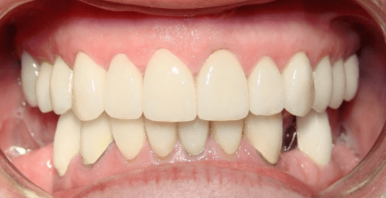
This patient had suffered a collapse of the bite due to wear, fillings, and bite adjustments. A comprehensive plan was devised, and since an opening of the bite was required, an orthotic was made to simulate the new chewing position. Once this was confirmed as comfortable, a wax simulation was again created to show the expected final result. At this point, the patient has had crowns on all viable remaining teeth and awaits implant replacement of the missing teeth. The ceramic was selected to be a whiter shade than the original.
(Shade Vita 3D 1M1, 14 Ivoclar e.max crowns, 6 PFM crowns. Teeth 36 and 37 extracted due to nonrestorable caries. Treatment plan calls for 34 and 36 implants to help support a tooth-and implant-supported PLD as lower right ridge insufficient to place implants and hip grafting refused. PFM’s on 43 and 44 splinted together with ERA attachment extending distally to avoid any show of denture clasps.)
Case 3
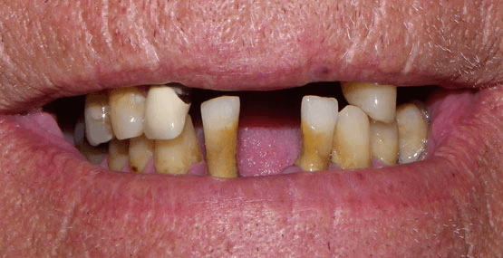
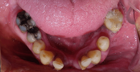
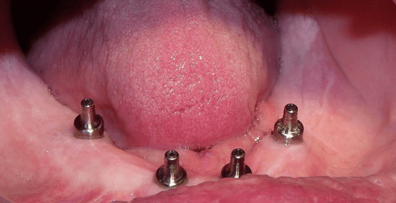
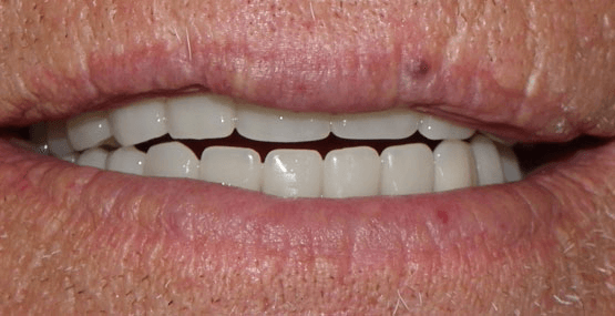
This was a patient referred to us by a local oral surgeon in order to coordinate surgical and prosthetic care. This patient was aware of multiple problems with his few remaining teeth and wanted to know his options.
After a detailed discussion, we advised him that the long-term prognosis of heroic attempts to save his remaining teeth would be poor, and instead recommended removal of all remaining teeth, the fabrication of an upper complete denture, and ultimately the placement of four lower implants to support a full lower “fixed-removable” denture (this is a denture that can be easily removed for cleaning, but is rock-solid in the mouth while being worn, so it doesn’t feel like a denture). Loose lower denture problems are over!
Surgery courtesy of Dr. Michael Jackson of Huronia Oral and Maxillofacial Surgery in Barrie, lab work courtesy of Shaw Laboratory Group in Toronto. Nobel Active implants, CAD/CAM milled titanium framework and abutments and framework courtesy of Panthera Dental Laboratory in Quebec City.
Case 4
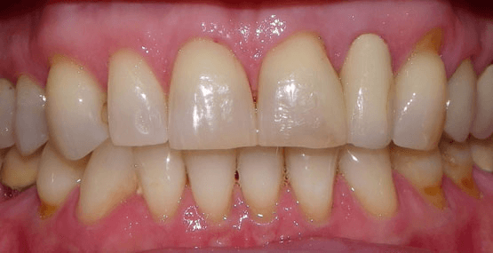
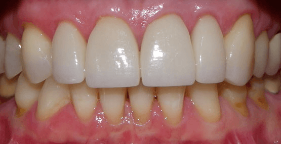
Here is another patient who had been coming regularly but just didn’t like the unevenness of her front teeth. Additionally, one of her front crowns had developed a crack that was beginning to stain. And, of course to complicate matters, she also had jaw fatigue which we felt was consistent with a lack of a single “home” position for her bite. This was a multi-step treatment that included:
- whitening
- reestablishment of a solid bite position using a Kois deprogrammer, leaf gauge, and subsequent bite equilibration
- crowns and veneers once the bite was comfortable
Why was it important to get the bite right? If we allowed her to continue chewing with multiple bites, the biting forces would be increased, and not only result in continued jaw pain, but would also increase the risk of porcelain fracture on the new crowns. This case was actually Dr. Wong’s presentation for the Kois Center Mentor examination.
(Whitening via KoR-Night 16% Carbamide peroxide gel. Kois deprogrammer courtesy of Caledonian Dental Laboratory in Barrie. e.max lithium disilicate veneers placed on 12,11,23 and crowns 21,22. Air abrasion of all teeth prior to bonding, acid etching with Bisco’s Uni-etch 32% H3PO4 (aq) with BAC. Veneers bonded with Calibra Adhesive resin cement using recommended protocol. Crowns cemented with 3M’s Unicem2.
Restorations courtesy of Image Dental Laboratory in Barrie.

