I don’t want X-rays. Or do I?: The Value of Seeing What You Can’t See
Posted: January 25, 2012
Last Modified: June 6, 2022
Periodically, we will see people who request not to have their dental x-rays (radiographs) taken. Sometimes this is for financial reasons. Sometimes it’s because they don’t think they “need” them. Maybe it’s the number of technologies out there that say that x-rays are no longer required. Or, maybe there are fears of radiation exposure. This post will highlight why this is often a case of “penny wise and pound foolish”.
First off, let us be clear: we subscribe to ALARA principles when it comes to radiation exposure (As Low As Reasonably Achievable). We want to get the information we need, but without exposing anyone to any more radiation than is necessary. We do this by performing a history and examination to guide us as to where and how (and even whether) to take the radiograph.
However, there is value to taking radiographs even when there is no pain or other symptom. We’ve already written here about why the absence of pain does not mean the absence of problems. If we detect an anomaly, it is wise to investigate before things get worse. Sometimes investigating involves the use of radiographs, and possibly even several radiographs at different angles.
So, what are the asymptomatic things that we might see that would lead to a radiograph? Of course, problems such as cavities (to determine the extent of decay) and gum disease (to determine the extent of bone loss), but there are many things that would spark some concern.
For children, this would include things like a high risk of decay (poor oral hygiene that would lead to cavities), teeth that are not coming in properly, missing teeth (in case they are impacted), orthodontic films, or teeth that should be lost by a certain age.We would also want to do follow-up films on traumatized teeth to see if they have died quietly.
For adults, there are also many reasons. These would include radiographs before procedures (for example, x-rays prior to extractions, root canals, crowns,bridges,implants and orthodontics, just to name a few), during procedures (mid-treatment films to check progress or healing), and after procedures (verification of a final result, e.g.: root canals). Similar to the case with children, any anomaly such as unexplained shifting of teeth or expansion of bony structures definitely requires a radiograph.
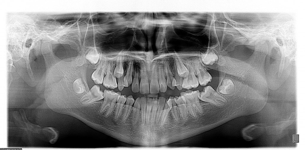
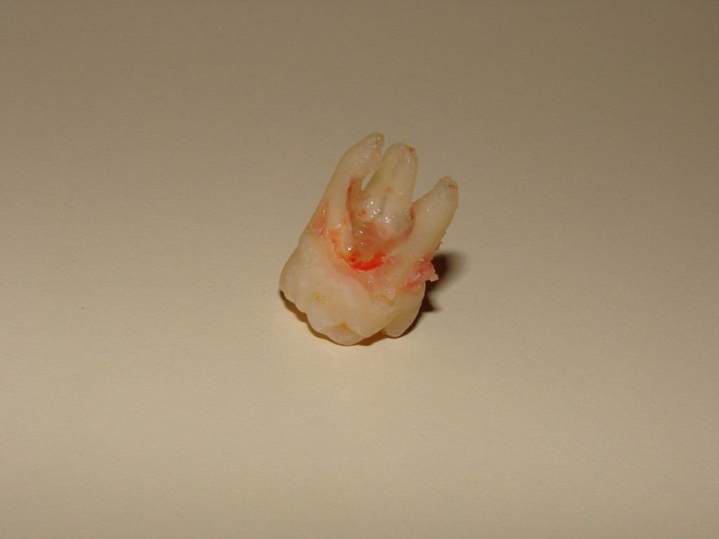
To illustrate the point, here is a case that presented to our office. This is a girl who had no reports of pain, but had some teeth that were not emerging properly. Some teeth were not present in the mouth long after they should have been visible. While it may be normal to assume some sort of routine orthodontic impaction problem, the x-ray (in this case, a panoramic x-ray) revealed significant problems with the upper first molars:


There is no method of saving the teeth with this kind of root resorption; extraction is the treatment of choice. With the molars out of the way, hopefully the second premolars previously stuck will emerge on their own.
This case serves to show where x-rays come in. Just because you can’t see it, or feel it, doesn’t mean it isn’t there.
Hope you enjoyed this post. For more topics of dental interest, keep visiting our Updates page for periodic information! Thanks for reading.
Please contact us if you would like us to have a look at your oral condition. We’d love to be your Barrie dentist.


